X-RAYS AND CHIROPRACTIC CARE
In order to provide the BEST QUALITY CARE, Dr. Quon utilizes a variety of diagnostic exams and tests to increase positive patient outcomes. One of these diagnostic tests includes spinal x-rays in office. New research has indicated the importance of using diagnostic radiology in order to provide patients with the BEST POSSIBLE OUTCOMES.
7 Reasons Why Radiography Used in Spine and Posture Rehabilitation Should Not Be Feared or Avoided
Evidence-based contemporary spinal rehabilitation often requires radiography. Use of radiography (X-rays or computed tomography scans) should not be feared, avoided, or have their exposures lessened to decrease patient dose possibly jeopardizing image quality. This is because all fears of radiation exposures from medical diagnostic imaging are based on complete fabrication of health risks based on an outdated, invalid linear model that has simply been propagated for decades. We present 7 main arguments for continued use of radiography for routine use in spinal rehabilitation: (1) the linear no-threshold model for radiation risk estimates is invalid for low-dose exposures; (2) low-dose radiation enhances health via the body’s adaptive response mechanisms (ie, radiation hormesis); (3) an X-ray with low-dose radiation only induces 1 one-millionth the amount of cellular damage as compared to breathing air for a day; (4) radiography is below inescapable natural annual background radiation levels; (5) radiophobia stems from unwarranted fears and false beliefs; (6) radiography use leads to better patient outcomes; (7) the risk to benefit ratio is always beneficial for routine radiography. Radiography is a safe imaging method for routine use in patient assessment, screening, diagnosis, and biomechanical analysis and for monitoring treatment progress in daily clinical practice.
Oakley, P. A., & Harrison, D. E. (2018). Radiophobia: 7 Reasons Why Radiography Used in Spine and Posture Rehabilitation Should Not Be Feared or Avoided. Dose-response : a publication of International Hormesis Society, 16(2), 1559325818781445. doi:10.1177/1559325818781445
X-Ray Imaging is Essential for Contemporary Chiropractic and Manual Therapy Spinal Rehabilitation: Radiography Increases Benefits and Reduces Risks
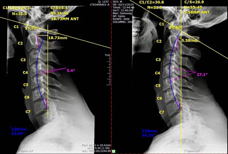
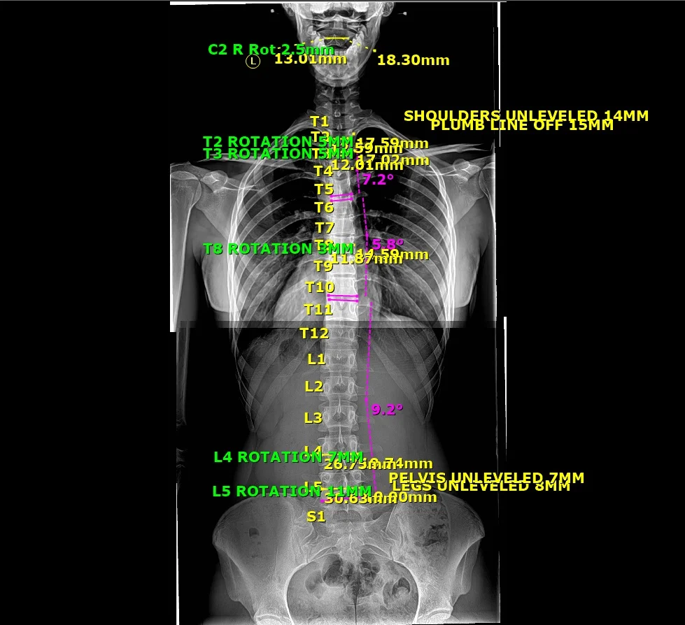
To remedy spine-related problems, assessments of X-ray images are essential to determine the spine and postural parameters. Chiropractic/manual therapy realignment of the structure of the spine can address a wide range of pain, muscle weakness, and functional impairments. Alternate methods to assess such spine problems are often indirect and do not reveal the root cause and could result in a significant misdiagnosis, leading to inappropriate treatment and harmful consequences for the patient. Radiography reveals the true condition and alignment of the spine; it eliminates guesswork. Contemporary approaches to spinal rehabilitation, guided by accurate imaging, have demonstrated superiority over primitive treatments. Unfortunately, there are well-meaning but misguided activists who advocate elimination or minimization of exposures in spine radiography. The radiation dose employed for a plain radiograph is very low, about 100 times below the threshold dose for harmful effects. Rather than increasing risk, such exposures would likely stimulate the patient’s own protection systems and result in beneficial health effects. Spine care guidelines need to be revised to reflect the potential benefits of modern treatments and the lack of health risks from low X-ray doses. This would encourage routine use of radiography in manual spine therapy, which differs from common pharmacologic pain relief practice.